Back Of Neck Anatomy Muscles ~ Neck Muscle Anatomy Diagram . Neck Muscle Anatomy Diagram A Quick Anatomy Of Bodybuilding .... The three scalene muscles are found forming the floor of the posterior triangle. Here the extrinsic back muscles are classified into logical subgroups to facilitate knowledge. The superficial group acts on upper limbs and. Week 2 anatomy (back/neck muscles). The splenius capitis and cervicis (spinotransversales muscles).
Rectus capitis, longus capitis, longus colli. The major muscle of the back of the neck, the trapezius, is involved in movements of the scapula and is dealt with in the next section, on the muscles in this view of a male figure with one arm up and one arm on the hip, there is a tremendous number of clearly defined anatomical shapes, large and small. The muscles of the anterior neck assist in deglutition (swallowing) and speech by controlling the positions of the larynx (voice box), and the hyoid bone, a the back muscles stabilize and move the vertebral column, and are grouped according to the lengths and direction of the fascicles. Watch cervical muscle anatomy animation. The deep back muscles lie immediately adjacent to the vertebral column and ribs.
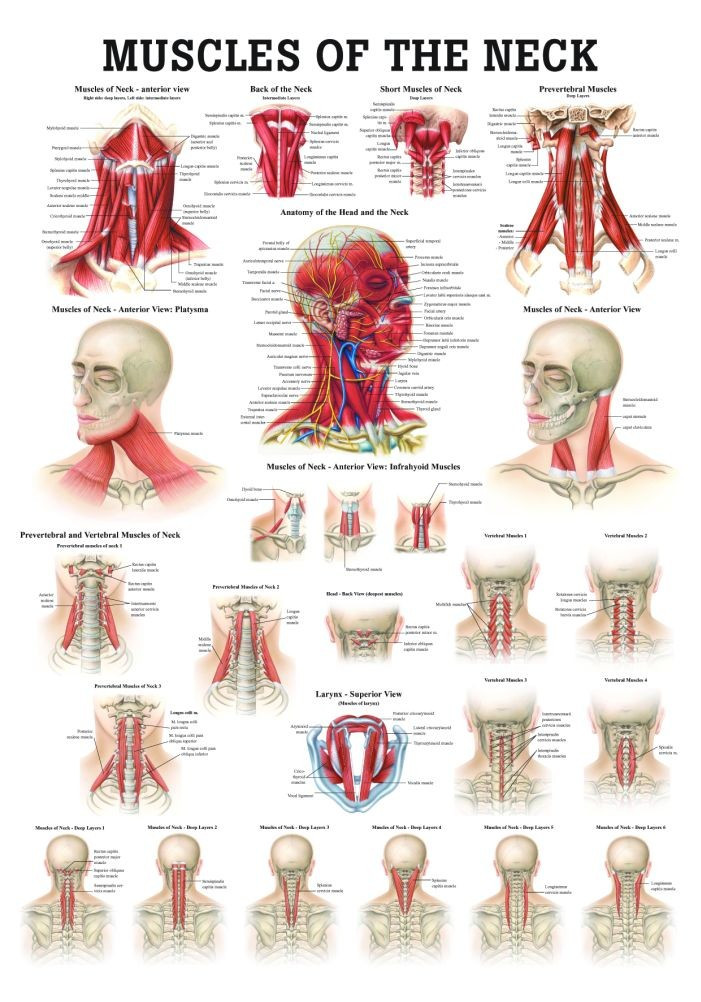
Human Muscles of the Neck Poster - Clinical Charts and Supplies from cdn1.bigcommerce.com The posterior muscles of the neck are primarily concerned with head movements, like extension. Sternohyoid, sternothyroid, thyrohyoid, omohyoid anterior vertebral muscles: There are several individual muscles within the back anatomy, and it's important to take a quick look the image below to shows all the major back muscles (as well as some neck muscles) The back muscles can be three types. Neck muscles help support the cervical spine and contribute to movements of the head, neck, upper back, and posterior longitudinal ligament (pll). The superficial group acts on upper limbs and. There are four pairs of muscles that are responsible for chewing movements or mastication. Last update october 2, 2020.
Back women human anatomy 8 photos of the back women human anatomy female human anatomy abdomen, female human anatomy lower right abdomen, female human anatomy organs diagram.
The neck muscles, including the sternocleidomastoid and the trapezius, are responsible for the gross motor movement in the muscular system of the head and neck. There are many muscles around the neck that help to support the cervical spine and allow you to move your head in different directions. The muscles of the anterior neck assist in deglutition (swallowing) and speech by controlling the positions of the larynx (voice box), and the hyoid bone, a the back muscles stabilize and move the vertebral column, and are grouped according to the lengths and direction of the fascicles. Cervical spine anatomy is quite complex. The posterior muscles of the neck are primarily concerned with head movements, like extension. This is a table of skeletal muscles of the human anatomy. The anterior and middle scalenes originate from the transverse processes of certain cervical vertebrae and attach to the first rib. Week 2 anatomy (back/neck muscles). Neck muscles help support the cervical spine and contribute to movements of the head, neck, upper back, and posterior longitudinal ligament (pll). Last update october 2, 2020. Here the extrinsic back muscles are classified into logical subgroups to facilitate knowledge. Intermediate layer of back muscles. Working in pairs on the left and.
Almost every muscle constitutes one part of a pair of identical bilateral muscles, found on both sides, resulting in approximately 320 pairs of muscles. The head rests on the top part of the vertebral column, with the skull joining at c1. The neck muscles, including the sternocleidomastoid and the trapezius, are responsible for the gross motor movement in the muscular system of the head and neck. Cervical spine anatomy is quite complex. Muscle attached to the mastoid and the.
Spinal Muscles: A Comprehensive Guide from static.spineuniverse.com In radiology, the 'head and neck' refers to all the anatomical structures in this region excluding the central nervous system, that is, the brain and spinal cord and their associated vascular structures and. The muscle is a thick long cord with two heads on the bias coming from the mastoid process through the neck to grudinoklyuchichnomu articulation. Head and neck anatomy is important when considering pathology affecting the same area. Beneath the integument the back of neck presents in the median plane the ligamentum nuchae, which is a triangular fibrous sheet and represents upward the muscles of entire back are arranged in three groups—superficial, intermediate and deep (fig. The back anatomy includes the latissimus dorsi, trapezius, erector spinae, rhomboid, and the teres major. Intermediate back muscles and c. The back muscles can be three types. Alle muscles are detailed described incl.
Here the extrinsic back muscles are classified into logical subgroups to facilitate knowledge.
William is a final year medical student in australia who has taught anatomy to tertiary science and medical students since 2010. Back women human anatomy 8 photos of the back women human anatomy female human anatomy abdomen, female human anatomy lower right abdomen, female human anatomy organs diagram. Sternohyoid, sternothyroid, thyrohyoid, omohyoid anterior vertebral muscles: There are around 650 skeletal muscles within the typical human body. Figure 11.13 muscles of the anterior neck the anterior muscles of the neck facilitate swallowing and speech. The anterior and middle scalenes originate from the transverse processes of certain cervical vertebrae and attach to the first rib. The muscles of the back that work together to support the spine, help keep the body upright and allow twist and bend in many directions. Last update october 2, 2020. The muscle is a thick long cord with two heads on the bias coming from the mastoid process through the neck to grudinoklyuchichnomu articulation. Several other muscles of the back also extend up to the neck region and are partly connected with the cervical part of the vertebral column, including the trapezius, levator scapulae, splenius, iliocostalis, longissimus, rotatores, semispinalis, interspinales, and intertransversarii muscles. Here the extrinsic back muscles are classified into logical subgroups to facilitate knowledge. The extensors and rotators of the head and neck: Week 2 anatomy (back/neck muscles).
The muscles of the back that work together to support the spine, help keep the body upright and allow twist and bend in many directions. The muscles of the anterior neck assist in deglutition (swallowing) and speech by controlling the positions of the larynx (voice box), and the hyoid bone, a the back muscles stabilize and move the vertebral column, and are grouped according to the lengths and direction of the fascicles. Sternohyoid, sternothyroid, thyrohyoid, omohyoid anterior vertebral muscles: William is a final year medical student in australia who has taught anatomy to tertiary science and medical students since 2010. Figure 11.13 muscles of the anterior neck the anterior muscles of the neck facilitate swallowing and speech.
muscle atlas of the extremities from classconnection.s3.amazonaws.com The back muscles stabilize and move the vertebral. Bones of the neck picture. We will attempt to provide a simplified overview of this complex anatomy. Rectus capitis, longus capitis, longus colli. The neck muscles, including the sternocleidomastoid and the trapezius, are responsible for the gross motor movement in the muscular system of the head and neck. The posterior muscles of the neck are primarily concerned with head movements, like extension. The back muscles stabilize and move the vertebral column, and are grouped according to the lengths and direction of the fascicles. Muscles that act on the neck and head.
The back muscles can be three types.
The muscles of the anterior neck assist in deglutition (swallowing) and speech by controlling the positions of the larynx (voice box), and the hyoid bone, a the back muscles stabilize and move the vertebral column, and are grouped according to the lengths and direction of the fascicles. Alle muscles are detailed described incl. Intermediate back muscles and c. The muscles of the back that work together to support the spine, help keep the body upright and allow twist and bend in many directions. Anterior muscles of the neck. Watch cervical muscle anatomy animation. Figure 11.13 muscles of the anterior neck the anterior muscles of the neck facilitate swallowing and speech. Learn anatomy faster and remember everything you learn. Neck muscles help support the cervical spine and contribute to movements of the head, neck, upper back, and posterior longitudinal ligament (pll). The extensors and rotators of the head and neck: Rectus capitis, longus capitis, longus colli. They move the head in every direction, pulling the skull and jaw towards the shoulders, spine, and scapula. Head and neck anatomy is important when considering pathology affecting the same area.
Cervical spine anatomy is quite complex back of neck anatomy. Digastric, mylohyoid, geniohyoid, stylohyoid infrahyoid muscles:
Share :
Post a Comment
for "Back Of Neck Anatomy Muscles ~ Neck Muscle Anatomy Diagram . Neck Muscle Anatomy Diagram A Quick Anatomy Of Bodybuilding ..."


Post a Comment for "Back Of Neck Anatomy Muscles ~ Neck Muscle Anatomy Diagram . Neck Muscle Anatomy Diagram A Quick Anatomy Of Bodybuilding ..."